3D Otoscope for enhanced diagnosis of middle ear-related issues
Eardrum elasticity, or lack of stiffness, is an important indicator for existing or impending middle ear-related issues such as early-stage middle ear inflammation, cholesteatoma, retraction pockets, and obstructive Eustachian tube dysfunction. Currently, Ear, Nose, and Throat (ENT) specialists do not have access to real-time, quantitative 3D shape information of the eardrum to supplement the two-dimensional qualitative view produced by regular otoscopy. The 3D otoscope addresses this need.

Situation before
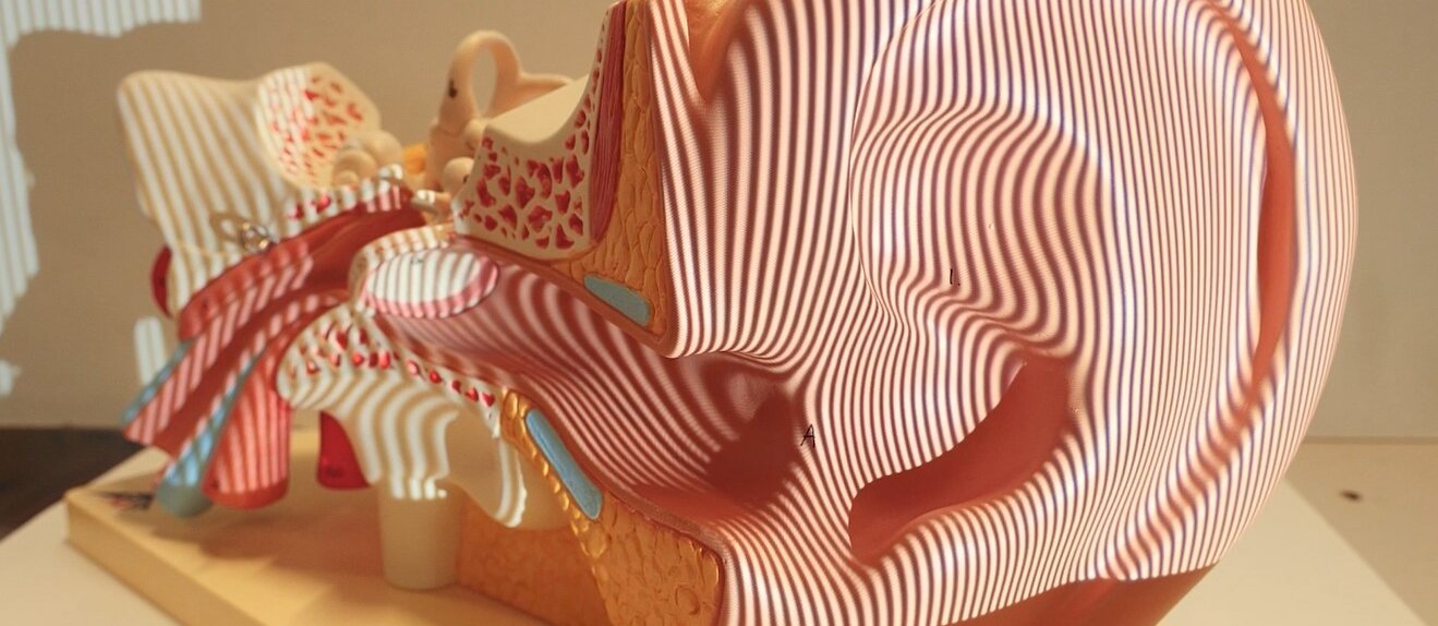
To assess the middle ear health in patients, ENT physicians typically use two types of diagnostic tests: classic 2D otoscopy and tympanometry. Classic otoscopy involves visually examining the eardrum to identify symptoms such as redness, scar tissue, retraction, bulging, and fluids. While some symptoms can be easily diagnosed based on these images, others may require further investigation to determine their severity. On the other hand, tympanometry is a quantitative method, but lacks spatial resolution. Tympanograms are created by sending pressure waves of a predetermined frequency range (sound) into the ear canal and measuring the amount of sound energy reflected from the eardrum. The resulting graphs can then be correlated with eardrum stiffness. However, in practice, these graphs often lead to false positive and false negative predictions of various pathologies.
In the current state of the art efforts to add a quantitative 3D measurement into an otoscopic device were unsuccessful due to practical size constraints associated with integrating a digital light projector in a small handheld device. Our innovation drastically simplifies the optical setup for handheld device integration.
Technology
We present a fully functional medical device (TRL 7) with a form factor similar to that of a regular otoscope. It has the capability of measuring the 3D shape of the human eardrum and its deformation in real-time (>30fps) at high resolutions (<50µm). The device and its operating principle are demonstrated in the clinical ENT setup and are protected for commercial exploitation through a patent granted in the EU and the US (WO2020152118A1).
We offer an industrially manufacturable handheld otoscopic device that is capable of measuring dynamic 3D height maps of human eardrums in-vivo and real-time. It works by projecting a single structured light pattern onto the eardrum surface and recording it with a high-speed camera. Both the optical imaging engine and the hardware setup of the device are designed to maximise depth accuracy when applied to uncoated human eardrums. By avoiding the need for coating, it can be considered non-invasive and classified as a class I medical device, significantly facilitating its market introduction. The main innovation (and part of the IP) is that our device operates using only a single projected pattern. This eliminates the need for bulky hardware such as triggering electronics, specialised lenses, cache memory to store multiple patterns, and significantly facilitates the device’s optical design.
By utilising a custom deep learning-based profilometry technique and a high-speed digital camera incorporated into a handheld device, we can measure the dynamic deformations of the eardrum and quantify volumetric displacements of the membrane in real time. This information will serve as a proxy for eardrum mobility and elasticity, providing valuable insights for physicians, and aligning with evidence-based medicine principles, enhancing diagnostic accuracy, and improving patient outcomes.
3D Otoscope for enhanced diagnosis of middle ear-related issues
Partner we search for
The potential revenues for a medical device like this are high, but so are the upfront costs of introducing it into a competitive medical market. Therefore, we are searching for a partner with a track record in bringing medical devices to market, an experienced marketing and sales department, and a broad distribution network.
About the researchers/research group
InViLab’s primary focus is on vision in all its forms, including camera calibration, multi- and hyperspectral imaging, 3D imaging, and thermal imaging. Medical device prototyping and the development of optical setups for challenging environments are at the core of our mission statement. Our team operates at the intersection of electromechanical prototyping, medical imaging, and real-time 3D imaging, including the continuous development of neural networks that enable single-shot profilometry.
More information
University of Antwerp
Valorisation Office
Middelheimlaan 1
2020 Antwerpen